Essential Tremor Treatment at Queen Square: Guidance for Patients and the People Close to Them
If you are living with essential tremor, or know someone who is, you might have come across a treatment called MR Guided Focused Ultrasound. You may have also stumbled upon something called FAT1 imaging. This guide explains these treatments in simple terms, how they work, and why the approach at Queen Square Imaging Centre is slightly different. No jargon here – just clear answers to the questions that really matter.
Living with Essential Tremor
Essential tremor means you have an uncontrollable shaking of your hands or arms when you try to do everyday things like write, eat or do up a button. Its not like it’s a restlessness or a tiredness, its happening when you’re actually trying to get on with your daily life.
For many people, it starts off gradually and gets worse with time. Its not uncommon for people to struggle with their work life and social life because of it, and it can even make everyday moments frustrating or embarrassing. And because its not always well understood, some people just go on living with it for years before finding out what treatment options are actually available.
Essential tremor is the most common movement disorder. More than 1 million people in the UK are living with it. People treated at Queen Square Imaging Centre had been living with the tremor for an average of 32 years before coming in for treatment
For a lot of people, medication is the first port of call. Some people find it helps, whereas some find that it bring no relief. For others, the side effects just get too much to handle – Fatigue, dizziness, not feeling 100 percent – even if the medication does stop the tremor, many find that the side effects make medication intolerable. If medication hasn’t worked for you or the person you care for then, don’t worry – there are other options out there.
What is MR Guided Focused Ultrasound (MRgFUS)?
MR Guided Focused Ultrasound (or MRgFUS for short) is a treatment that enables you to reduce the impact of your essential tremor without having to go under the knife. No cuts, no stitches and no implants are required. Nothing goes inside your body
So, how does it work? The treatment uses focused beams of sound energy. Hundreds of beams are fired through your skull and focused on one tiny spot deep in the brain. At that spot, the combined energy builds up enough heat to treat the bit of the brain that’s causing the tremor. Everywhere else gets a tiny fraction of that energy and is totally fine
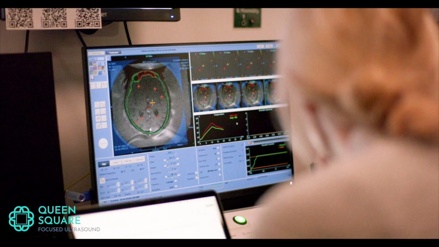
The whole thing happens inside an MRI scanner, which is doing two jobs at once: creating the images the clinical team need to make sure they’re in the right spot, while also tracking the temperature in real time so they can see exactly what’s happening and make any adjustments as they go.
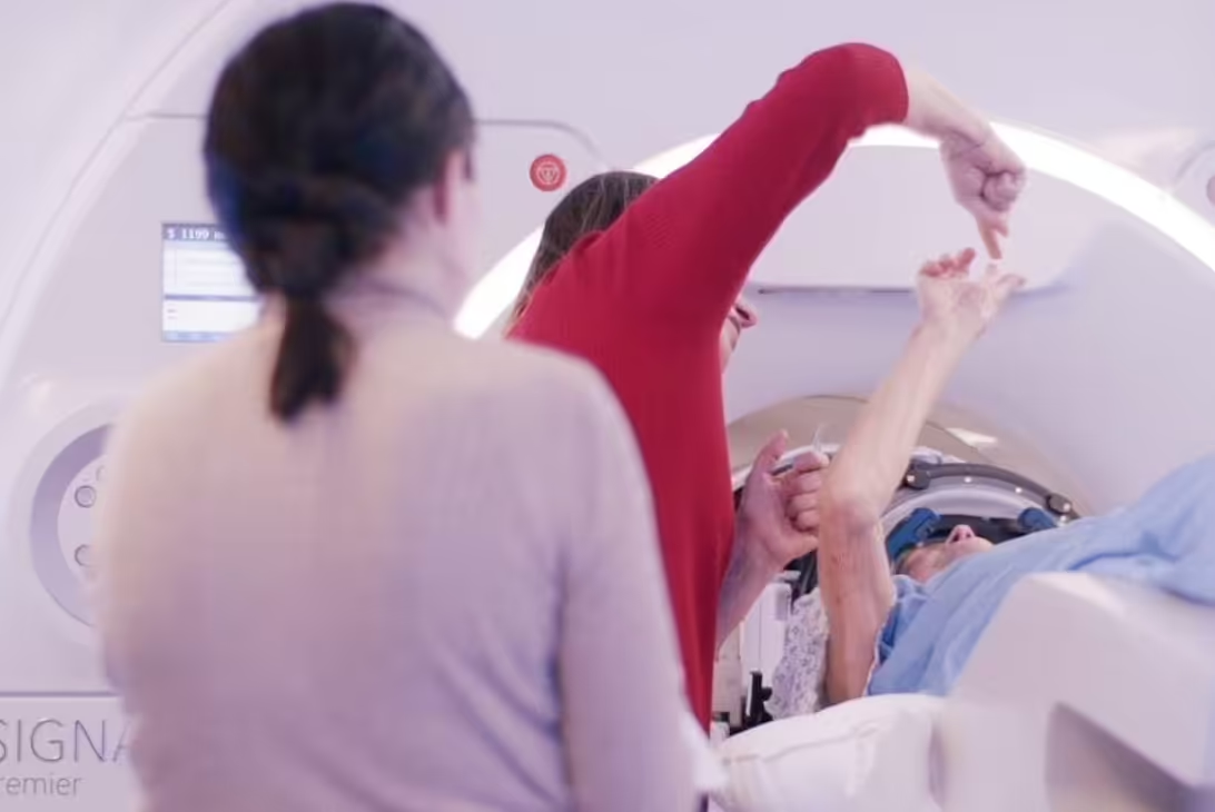
You stay awake the whole time, no anaesthetic needed. The clinical team will talk to you, test your tremor and check in on how you’re feeling after each little blast of sound energy. Your response guides the treatment as it happens.
Is MRgFUS painful?
Most people experience some mild sensations during the treatment, including a temporary feeling of warmth or pressure, occasional dizziness or nausea, and sometimes a brief tilting sensation. These are common and generally short-lived. Many people find them manageable. The clinical team monitors you closely throughout and can pause or adjust the treatment at any point.
Why Getting the Right Target is So Important
The tremor is caused by a tiny part of the brain called the Ventral Intermediate Nucleus – the Vim for short. Its deep in the brain and the circuits that make the shaking happen all go through it. So, if you can sort out that specific bit of the brain, you can sort out the tremor.
The problem is that the Vim is tiny – and standard MRI scans just can’t show it well enough to target it directly.
Traditionally, teams have worked around this by using maps of the brain, based on lots of averages from lots of different people, to try and work out where the Vim might be. Whilst this technique does work, every brain is a bit different. What might be average for lots of people might not be right for you.
When the initial target is a bit off, its not the end of the world, but it does make things a bit tricky. The team might need to do a few extra bursts of ultrasound to find and test out the right spot. Not only is that a bit more work for both you and the treatment team, but research has also shown that the risk of side effects goes up when more bursts of ultrasound are used. Research shows that in a lot of standard MRgFUS programmes, the initial aim actually has to be corrected more often than not.
FAT1 imaging was designed to solve that problem by making the Vim visible, so the team can accurately target it in your brain, rather than just using an estimate of where it might be.
FAT1 Imaging – The Breakthrough in Brain Scanning
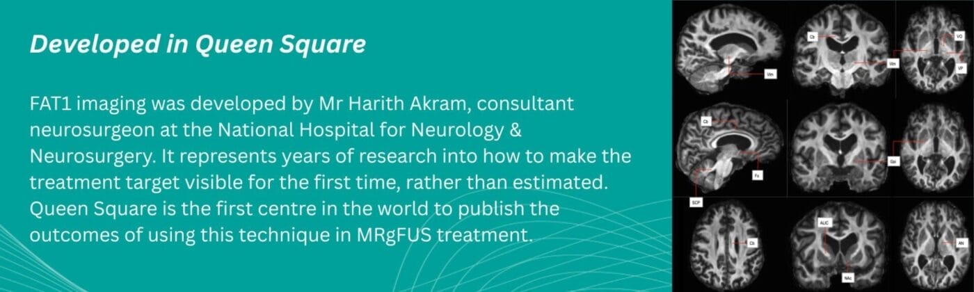
FAT1 imaging is a specially developed type of brain scan, created by the clinical team at Queen Square Imaging Centre and the National Hospital for Neurology and Neurosurgery. It combines two different MRI scans into one image. When these two types of scan are merged, something becomes visible that neither could show on its own: the Vim, clearly enough to target it directly.
One of the two scans tracks the movement of water molecules along the nerve fibres in the brain. In areas where fibres run in a tight, organised way, the water follows a consistent path. This reveals the underlying structure of the brain in a way that ordinary MRI cannot. When that information is combined with a detailed structural brain scan, the result is an image in which the Vim can be seen as a distinct structure.
Think of it this way. Standard MRI is like looking at a city on a satellite image, but without street names or labels. You know you can see the buildings & roads, but you couldn’t pinpoint a specific address without some educated guessing. FAT1 imaging adds the street labels – the target is no longer an estimate. It’s right there in front of you.
What Happens in Queen Square: Step by Step guide to MRgFUS
Before your procedure
A few weeks before the treatment day, you will have a detailed MRI scan. This takes longer than a standard scan because the images need to be highly detailed. These scans are then used to generate your personal FAT1 image, which shows the treatment team exactly where your Vim is located in your own brain.
Your FAT1 image is then loaded into the treatment planning system alongside a CT scan of your skull. The team uses these together to plan your treatment precisely, mapping exactly where the sound energy should be directed on the day.
On the day of your procedure
You will arrive at Queen Square Imaging Centre and a frame will be gently fitted to your head to keep it still during the treatment. You will then be positioned inside the MRI scanner with a water-filled cap fitted around your head. This is the device through which the focused ultrasound waves are delivered.
The clinical team will be with you throughout. A movement disorders neurologist will assess your tremor and check how you are feeling after every pulse of sound energy. Because you are awake, your response is part of how the treatment is guided.
The team will start with a small number of low-power pulses to check that everything is accurately positioned. When they are satisfied with the aim, the therapeutic pulses begin. These heat the treatment area to the temperature needed to settle the tremor-causing tissue. Each pulse lasts only about 12 seconds.
Because FAT1 imaging means the initial target is set with confidence, the number of pulses needed is lower than in standard programmes, and the overall treatment time is shorter. At Queen Square Imaging Centre, the active treatment phase takes an average of around 35 minutes.
After the procedure
Many patients notice an improvement in their tremor during the procedure itself, sometimes immediately after the first therapeutic pulse. You will be monitored closely before going home.
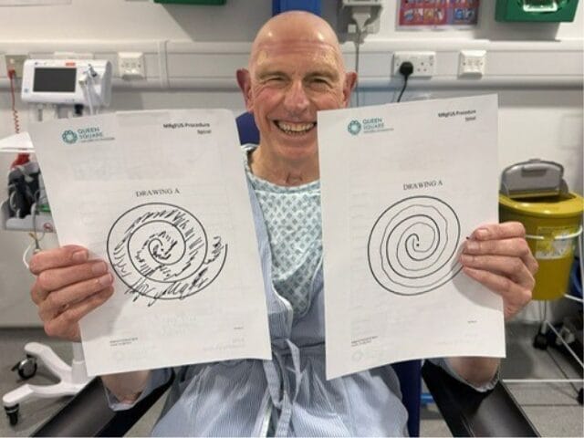
There may be some mild side effects in the days after treatment, including temporary changes in balance or coordination. The clinical team will advise you on what to expect and what to watch out for. All patients are advised to use a walking stick for the first week after the procedure as a precaution.
What Results Can Patients Expect?
The first published study of FAT1 imaging in MRgFUS treatment, conducted at Queen Square Imaging Centre and the National Hospital for Neurology and Neurosurgery and published in a peer-reviewed medical journal in 2025, followed 14 patients for 12 months after treatment. The results at 12 months were as follows.
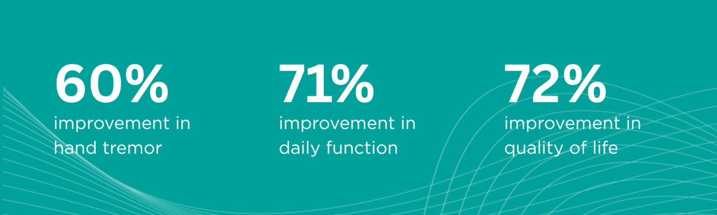
All 14 patients reported improvement in quality of life. None reported side effects at 12 months that affected their ability to function day to day.
The quality of life improvement of 72% is also notably higher than figures reported in previous MRgFUS studies, which typically reported improvements of around 45 to 46%. The study team attributes this partly to the greater precision of FAT1-guided targeting.
“FAT1 imaging is a game-changer. By improving the visibility of the brain structures we need to target, we can deliver this incisionless treatment with greater accuracy, faster results and fewer side effects, making a meaningful difference to patients’ lives.”
Mr Harith Akram, Consultant Neurosurgeon, National Hospital for Neurology & Neurosurgery
Is This Treatment Right for Me?
MR Guided Focused Ultrasound is not suitable for everyone. It is offered to patients with essential tremor whose symptoms have not been adequately controlled by medication. All patients are assessed individually by a specialist team before any treatment is recommended.
There are some factors that affect whether someone is suitable for the procedure, including the density of the skull bones, which affects how well the sound waves can be focused. This is assessed during the initial consultation using a CT scan.
The best way to find out whether you are eligible is to speak to your GP about a referral to Queen Square Imaging Centre. You can also visit queensquare.com to read more about the treatment, the assessment process and what to expect.
Next Steps: Here’s What to Do
Contact us today to begin your journey. Whether you want further information, or would like to discuss your own assessment, our team of treatment coordinators are on hand to assist.
MORE NEWS