New imaging technique could improve outcomes for essential tremor patients
A new study by world-leading neurosurgeons from Queen Square Imaging Centre in London has unveiled a breakthrough imaging technique that significantly enhances the precision and outcomes of MR-guided focused ultrasound (MRgFUS) – a revolutionary, non-invasive treatment for essential tremor, the most common movement disorder in the world.
Affecting over a million people in the UK and millions more worldwide, essential tremor causes uncontrollable shaking, typically in the hands, severely impacting an individual’s work, independence and mental wellbeing. While medication is often the first line of treatment, it fails to help many patients long-term, leading to the need for more advanced interventions.
Now, a new technique using FAT1 imaging – a sophisticated MRI approach that fuses multiple scan types – is offering new hope to patients who previously faced invasive surgeries or limited treatment options.
The study published in The BMJ Neurology Open marks the first clinical use of FAT1 imaging to guide focused ultrasound treatment for essential tremor. Traditionally, clinicians have relied on generalised brain maps to estimate the location of the target area deep within the brain – the Ventral Intermediate Nucleus (Vim) – which is extremely small and hard to visualise on standard MRI scans.

Image 2 shows an example of the FAT-1 images used by neurosurgeons at Queen Square Imaging Centre.
FAT1 imaging overcomes this by giving surgeons a clear, direct view of the individual patient’s Vim, enabling treatment to be far more precise.
“FAT1 imaging is a game-changer,” said Mr Harith Akram, a consultant neurosurgeon at Queen Square Imaging who developed the technique himself and led the research. “By improving the visibility of the brain structures we need to target, we can deliver this non-invasive treatment with greater accuracy, faster results and fewer side effects, making a meaningful difference to patients’ lives.”
Study highlights: strong results for patients
- 14 patients with essential tremor were treated using MRgFUS guided by FAT1 imaging
- Tremor improved by an average of 60% at 12-month follow-up
- Quality of life significantly enhanced in all patients
- Side effects were milder and shorter-lasting, with better avoidance of nearby brain regions
- Procedures were faster, required less energy, and achieved accurate targeting on the first attempt in every case
A non-invasive future for tremor treatment
Unlike traditional treatments such as Deep Brain Stimulation (DBS) or Radiofrequency Ablation (RFA), MRgFUS is completely non-invasive and incisionless. It works by focusing sound waves to gently heat and destroy the tremor-causing area of the brain, all carried out under real-time MRI guidance.
With no surgery, no implants and a quicker recovery, MRgFUS is increasingly being seen as the treatment of choice for eligible patients who do not respond to medication.
“This study represents the future of tremor treatment,” added Mr Akram. “With new techniques like FAT1 imaging, we’re able to personalise care in a way that is safer, smarter and more effective and ultimately give patients a better chance at regaining control and improving their quality of life.”
Patients should always seek the advice from a professional and discuss their condition with a neurologist or movement disorder specialist to see if it’s suitable for their individual case.

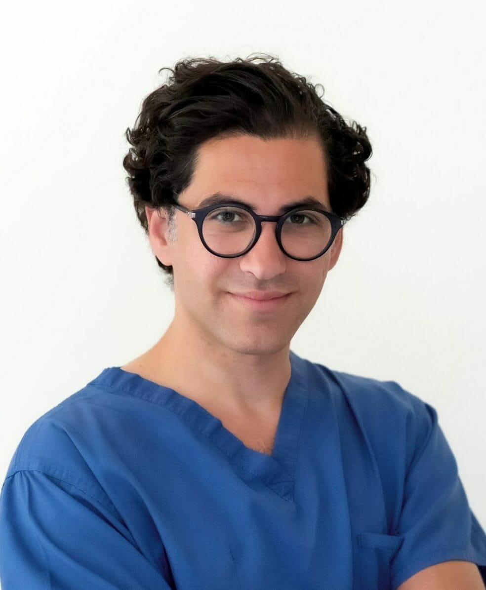
Mr Harith Akram, a consultant neurosurgeon at Queen Square Imaging Centre, who developed the FAT-1 imaging technique and led the research.
Mr Harith Akram is a consultant neurosurgeon and part of the world-leading team at Queen Square Imaging Centre. Mr Akram is a pioneer of connectivity guided thalamic guided surgery for tremor and the Editor-in-chief of the World Society of Stereotactic and Functional Neurosurgery (WSSFNS) newsletter.
MORE NEWS


