
New directions in the assessment of mild traumatic brain injury
Traumatic Brain Injury – A leading health concern
Traumatic Brain Injury (TBI) results in around 1.4 million hospital visits in England and Wales and stands as the leading cause of mortality among individuals under 40 years old. Of these cases, approximately 10% are severe, 10% are moderate, and 80% are classified as mild. The true incidence rate of mild Traumatic Brain Injury (mTBI) is challenging to ascertain as not all cases are evaluated or reported at a hospital.
Challenges in the diagnosis of mild traumatic brain injury
The shearing forces acting on the brain during trauma can lead to axonal disruption, specifically diffuse axonal injury, which may explain why 10-20% of mTBI patients continue to experience symptoms for extended periods post-injury. Diagnosis of mTBI has traditionally relied on the Glasgow Coma Scale (GCS), yet this method does not offer detailed information about the location of the injury, damage, or the potential impact on the individual, making the diagnostic process complex and susceptible to both over and under diagnosis depending on the criteria used by the physician.
Advancements in neuroimaging for diagnosing mild traumatic brain injury
Common symptoms typically fall into three categories: physical, cognitive, and behavioural. Due to the absence of definitive biomarkers and the wide array of diagnostic criteria, clinicians often approach mTBI diagnosis through a combination of clinical tests and neuroimaging scans, such as MRI. While MRI has become essential in assessing brain injuries, including mTBI, routine post-injury scans often fail to correlate with the severity and long-term cognitive outcomes in mTBI cases. Consequently, recent research has been directed towards identifying a neuroimaging technique that can accurately diagnose mTBI and align with its clinical outcomes, with diffusion tensor imaging (DTI) emerging as a promising advanced MRI technique.
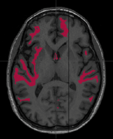
DTI, being highly sensitive to microstructural injuries not visible on conventional MRI, could serve as a non-invasive neuroimaging tool for diagnosing mTBI, providing surrogate biomarkers for clinical performance scores, objective documentation of diagnosis, tracking changes over time, biomarkers for clinical trials, and aiding clinicians in ruling out similar diseases with overlapping symptoms that may delay treatment.
Conclusion: Towards better diagnosis and management of mTBI
The complexities surrounding the diagnosis and management of mild Traumatic Brain Injury (mTBI) highlight the need for more accurate and reliable diagnostic tools. While traditional methods such as the Glasgow Coma Scale and standard MRI scans have been pivotal, their limitations underscore the necessity for advanced techniques. Diffusion Tensor Imaging (DTI) presents a promising solution, offering greater sensitivity to microstructural brain injuries and the potential to provide more detailed insights into the impact of mTBI. As research progresses, the integration of such advanced neuroimaging techniques could revolutionize the way mTBI is diagnosed and managed, ultimately improving outcomes for patients and reducing the long-term burden of this common yet often underreported injury.

MORE NEWS

