
Imaging Stroke: How MRI scanning has revolutionised diagnosis and Treatment
MRI (Magnetic Resonance Imaging) has revolutionised the diagnosis and management of stroke, becoming an indispensable tool in current diagnostic, treatment and rehabilitation pathways. In the intricate landscape of neurological disorders, stroke stands out as an urgent and often life-altering event. With its unique ability to provide detailed anatomical images and valuable quantitative information about the physiological process taking place within the brain, MRI scans offer unparalleled insights into the brain’s structure and function, making the imaging technique pivotal in stroke diagnosis, treatment planning, and prognosis.
At the forefront of this medical frontier stands the Queen Square Imaging Centre, a beacon of excellence in neuroimaging for over four decades. Located in the heart of London and working in partnership with the renowned National Hospital for Neurology and Neurosurgery, QSIC is highly regarded for its use of cutting-edge technology and radiological expertise, dedicated to providing unparalleled patient care and support.

In the context of stroke, MRI offers crucial advantages over other imaging modalities. Its ability to capture detailed images of the brain’s anatomy and blood flow allows clinicians to pinpoint the location, extent, and type of stroke with remarkable precision. More recently, advanced MRI techniques have enabled early detection of stroke-related changes, facilitating prompt intervention and minimising long-term damage. Owing to decades of experience and development, QSIC now routinely offers a combination of the following techniques to reveal further insight:
Diffusion-Weighted Imaging (DWI)
When a stroke occurs, brain cells are deprived of oxygen and start to die. DWI can contribute valuable information by detecting these changes very early on by measuring the movement of water molecules in the brain. In a healthy brain, water molecules move around freely. However, during a stroke, this movement is restricted due to cell damage. DWI picks up on these restricted movements, allowing the radiologist to pinpoint the affected areas quickly. This early detection is vital because it helps stroke clinicians make decisions about treatment options, potentially minimising the risk of long-term damage from the stroke.

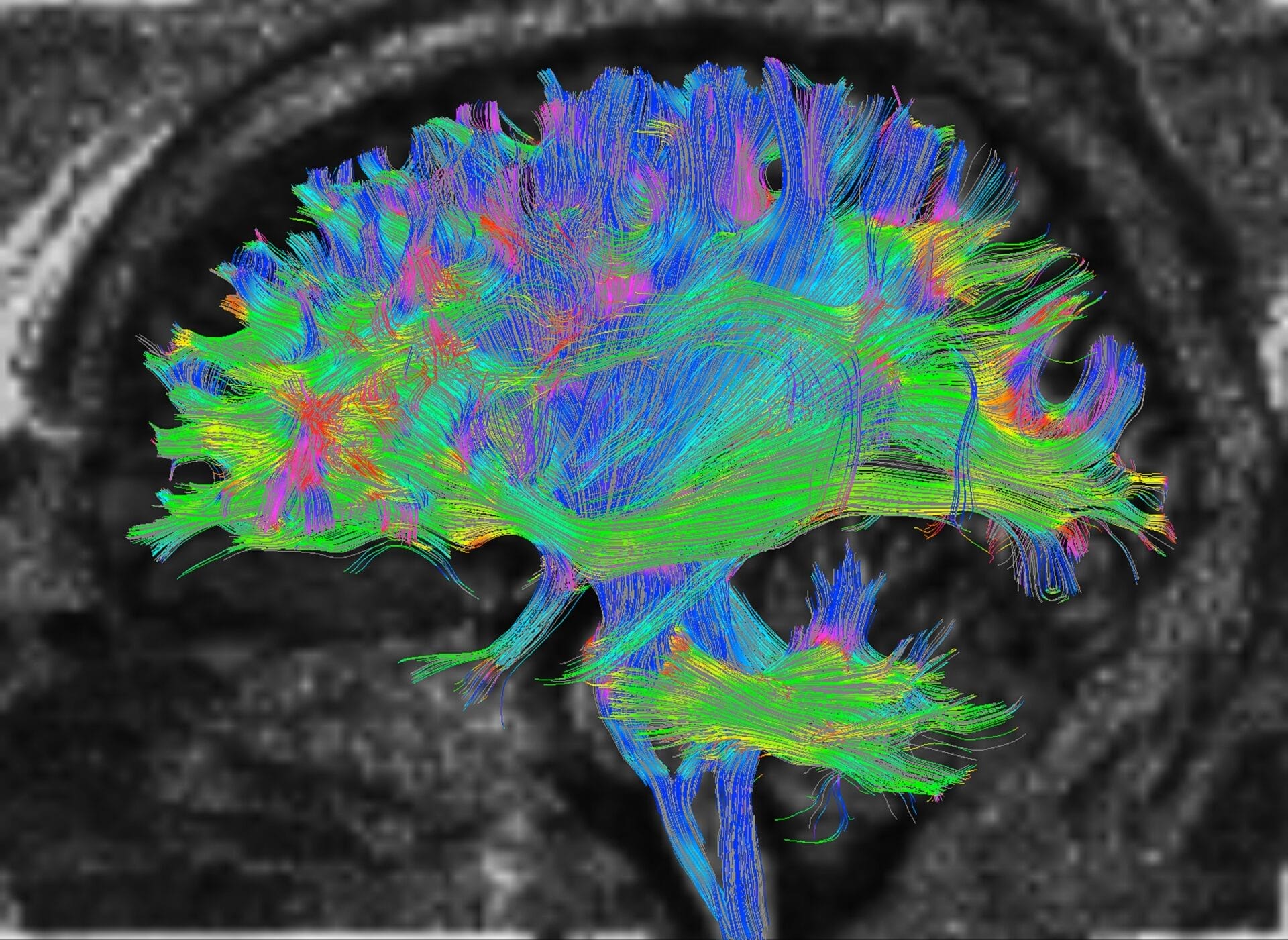
Diffusion Tractography (DTI)
In the context of stroke, DTI offers detailed insights into the integrity of the brain’s white matter tracts, which are crucial for transmitting signals between different brain regions. When a stroke occurs, these pathways can be disrupted, leading to various neurological deficits. DTI helps in visualizing these disruptions, aiding clinicians in assessing the extent of damage caused by the stroke and predicting functional outcomes. By mapping the affected white matter tracts, DTI helps in personalised treatment planning and rehabilitation strategies, ultimately improving the patient’s recovery and quality of life post-stroke.
Susceptibility Weighted Imaging (SWI)
Stroke is often associated primarily with a blockage of a blood vessel causing an interruption to blood flow to the brain. However, haemorrhagic strokes (bleeding within the brain tissue) are another common. SWI is particularly sensitive to blood products due to the magnetic properties of iron rich blood. SWI enables the visualisation of even the smallest amounts of bleeding in the brain, enabling exact characterisation of the type and extent of stroke, guiding treatment decisions, and predicting patient outcomes.

Perfusion Weighted Imaging
Perfusion MRI is a key tool in stroke assessment, providing valuable information about blood flow to the brain tissue. During a stroke, blood flow is disrupted, leading to ischemia (inadequate blood supply) in affected areas. Perfusion imaging in the case of acute stroke measures blood flow, volume, and transit time, helping clinicians to find regions of reduced perfusion, which may indicate areas at risk of irreversible damage, or penumbra (potentially salvageable tissue surrounding the core infarct). This information guides treatment decisions, such as the administration of thrombolytic therapy or mechanical thrombectomy, aiming to restore blood flow and minimise brain injury. Perfusion MRI also aids in predicting patient outcomes and assessing treatment response, helping personalised stroke management strategies for improved patient care and recovery.

Conclusion
MRI has emerged as a cornerstone of stroke management, offering unparalleled insights into the brain’s inner workings. With its ability to visualise stroke-related changes with exquisite detail, MRI guides diagnosis, treatment, and research, shaping the landscape of stroke care for generations to come.
