Colloid Cysts: A Comprehensive Guide to Symptoms, Diagnosis, Imaging, and Treatment
Colloid cysts are rare, benign, fluid-filled growths that form in the brain. While they account for only about 1-3% of all brain tumours, their location and potential to cause significant symptoms make them a critical condition to recognise and manage effectively. This article explores what colloid cysts are, the symptoms they can cause, their appearance on imaging studies, current treatments available, and strategies for management.
What Are Colloid Cysts?
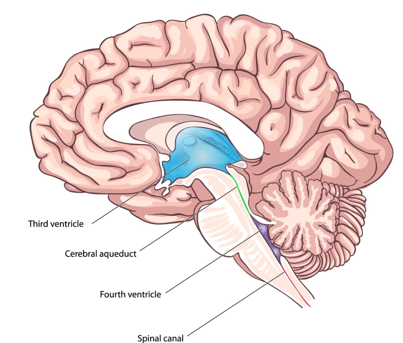
Colloid cysts are small, sac-like structures with a thin epithelial lining and typically filled with a gelatinous material often containing mucin (a family of proteins found in mucus), old blood and cholesterol. These cysts most commonly occur in the third ventricle of the brain, a central cavity that is part of the normal circulation of cerebrospinal fluid (CSF) around the central nervous system. Due to their position, colloid cysts can sometimes obstruct the normal flow of CSF, leading to a condition called hydrocephalus (fluid build-up in the brain). Understanding how colloid cysts can affect both the brain and spinal cord is crucial for effective surgical and non-surgical patient management.
While the exact cause of colloid cysts is unknown, they are believed to arise during fetal development and are often detected in adulthood as either an incidental finding, or when their size starts to cause symptoms, typically between the ages of 20 and 50.
Characteristics of Colloid Cysts
Colloid cysts are rare, benign brain tumours that develop in the fluid-filled regions of the brain known as ventricles. Most commonly, these cysts are found in the third ventricle, which is situated deep within the brain.

Unlike the type of brain tumours, we may more often think about, colloid cysts are filled with a proteinaceous fluid and are typically attached to the roof of the third ventricle or the choroid plexus (a structure which produces CSF). These cysts account for approximately 15 to 20 percent of all masses that arise within the ventricles, making them a notable, though uncommon, type of brain tumour.
Symptoms of a Colloid Cyst
Symptoms of colloid cysts can vary widely, depending on the size and precise location of the cyst and the extent to which it causes a CSF blockage. Some individuals may remain asymptomatic, with the cyst discovered incidentally during medical imaging for unrelated reasons. Others may experience significant symptoms that lead to investigation and diagnosis, including:
- Headaches: These are often severe and positional, worsening when lying down.
- Nausea and vomiting: Resulting from increased intracranial pressure.
- Blurred or double vision: This can be secondary to pressure exerted on surrounding brain structures.
- Cognitive or memory problems: In rare cases, pressure exerted by a cyst can affect brain function.
- Sudden neurological decline: In extreme cases, acute blockage of CSF can lead to rapid onset symptoms, including unconsciousness or sudden death, though this is rare.
Imaging Appearance on Magnetic Resonance Imaging and CT
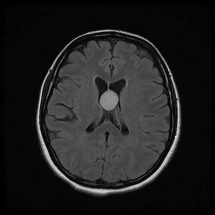
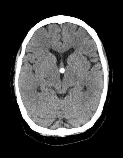
Colloid cysts are often identified through Magnetic Resonance Imaging (MRI) or Computed Tomography (CT or CAT) imaging:
On MRI (Magnetic Resonance Imaging):
- Cysts appear as well-defined, round masses in the third ventricle.
- Signal intensity, which is used to define and characterise a lesion on MRI, varies on the different types of imaging that may be acquired during the MRI scan. A combination of images known as T1- and T2-weighted images would be performed, and the cyst’s appearance will vary on each, depending on the cyst’s fluid composition.
- Cysts may enhance following the injection of an MRI contrast dye in some cases.

On CT (Computed Tomography):
- Cysts may appear as hyperdense lesions (meaning denser or ‘lighter’) or isodense lesions (meaning the same density) when compared to adjacent normal brain tissue.
- Colloid Cysts may occasionally show areas of calcification in some cases, which can often help in distinguishing them from other lesions.

High quality imaging using MRI or CT is essential, not only for diagnosis but also for monitoring cyst size and for potential complications like hydrocephalus. It is not uncommon for patients with known colloid cysts to have repeat imaging at set intervals so that the cyst can be closely monitored over time.
How are Colloid Cysts treated?
The treatment approach for a colloid cyst will be personalised to each patient, and will depend on the size of the cyst, the presence and severity of symptoms, and the degree of CSF obstruction. Treatment may include one, or a combination of the following:
- Observation and Monitoring:
- Asymptomatic cysts with no hydrocephalus may not require immediate intervention.
- Regular imaging (using MRI or CT) will be used to help monitor growth or changes over time. This will then be crucial to informing decisions about more active treatment in the future,
- Surgical Removal:
- For symptomatic cysts or those causing hydrocephalus, surgery is the primary treatment.
- Minimally invasive endoscopic surgery is increasingly preferred, offering a shorter recovery time and lower complication rates compared to open craniotomy.
- Microsurgical resection may be necessary for larger or more complex cysts.
- CSF Diversion Procedures:
- For patients unable to undergo cyst removal, a ventriculoperitoneal (VP) shunt or endoscopic third ventriculostomy (ETV) can alleviate hydrocephalus by re-routing CSF flow via a synthetic route (a shunt) or by creating a new exit in the patient’s own anatomy (ventriculostomy).
Complications and Risks
Colloid cysts can lead to a variety of complications and risks, primarily due to their potential to obstruct the flow of cerebrospinal fluid (CSF). This obstruction can result in hydrocephalus, a condition characterised by excessive collection of CSF within the brain, leading to increased intracranial pressure. Common complications include:
- Hydrocephalus: The blockage of CSF flow can cause fluid build-up, leading to increased pressure within the brain.
- Headaches: Often severe, these headaches are typically most intense in the morning upon waking.
- Nausea and Vomiting: These symptoms are particularly prevalent if the cyst is large or causing significant hydrocephalus.
- Blurry Vision: Pressure on the optic nerve can result in visual disturbances.
- Gait Abnormality: Changes in walking patterns, along with personality changes, memory loss, double vision (diplopia), swelling of the optic disc (papilledema), and sudden falls (drop attacks), can occur.
Prognosis and Outlook
The prognosis and outlook for individuals with colloid cysts largely depend on the cyst’s size, location, and the severity of symptoms. Small, asymptomatic cysts may only require close observation with periodic imaging tests, such as MRI scans, to monitor for any changes. If the cyst is large or symptomatic, surgical treatment is often necessary. Surgical options include craniotomy, endoscopic craniotomy, or shunt placement to alleviate symptoms and prevent complications.
With appropriate surgical treatment, the prognosis for colloid cysts is generally favourable, and most patients can expect to make a full recovery. However, there is a possibility of recurrence, necessitating ongoing monitoring and, in some cases, additional treatment. By adhering to a comprehensive treatment plan and regular follow-ups, patients can effectively manage their condition and maintain a good quality of life.
Management Advice for Patients with Colloid Cysts and Cerebrospinal Fluid Issues
- Stay Informed: Understanding your condition and the potential risk of a colloid cyst is crucial.
- Monitor Regularly: If the cyst is asymptomatic, follow your doctor’s recommendations for periodic imaging.
- Know the Signs of Increased Pressure: Symptoms like worsening headaches, nausea, or sudden confusion may indicate hydrocephalus and require immediate medical attention.
- Discuss Treatment Options: If surgery is recommended, explore the pros and cons of endoscopic versus microsurgical techniques with your neurosurgeon.
- Lifestyle Considerations: Maintaining a healthy lifestyle and managing stress can support overall brain health, though these factors won’t directly influence the cyst itself.
Conclusion
Colloid cysts, while rare, can have significant implications for health due to their potential to obstruct CSF flow and cause serious symptoms. Early detection through imaging and careful monitoring are essential, and treatment options have advanced to offer safer, less invasive procedures. By working closely with medical professionals and staying vigilant about symptoms, patients can effectively manage this condition and maintain a high quality of life.


