CaRi-Heart – A new tool for providing personalised assessment of the risk of heart attack.
What is Coronary Artery Disease?
Coronary artery disease (CAD) is a leading cause of mortality worldwide, affecting millions of individuals each year. It occurs when plaque (a build-up of fatty material) builds up in the blood vessels that supply blood to the heart muscle. This leads to reduced blood flow, potentially causing chest pain, heart attacks, or other cardiovascular complications. Atherosclerosis, the disease process that causes the build of plaque in the coronary artery blood vessels is an inflammatory disease, as is the process which drives the rupture of unstable plaques which leads to heart attack.
Early detection and management of CAD are crucial for preventing serious health issues.

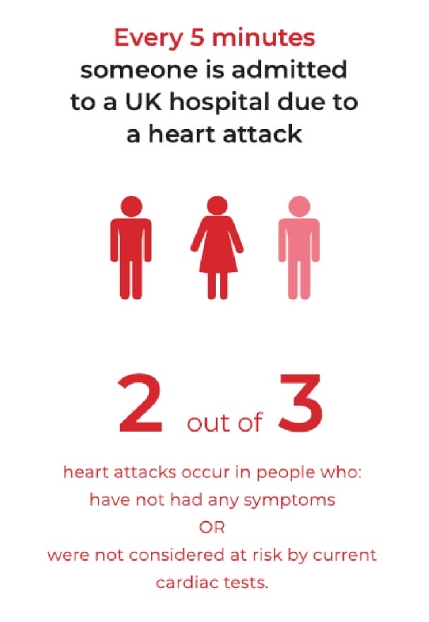
Source: Protect Your Heart – Caristo Diagnostics 2021
The role of CT Coronary Angiography in CAD
The 2016 NICE guidelines recommend that CT Coronary Angiography (CTCA) should be used as the first-line investigation for patients with stable chest pain and a suspicion of CAD. A relatively fast, easy and non-invasive imaging test, CTCA aims to detect the narrowing in coronary arteries which may result in reduced blood flow and damage to the heart muscle (ischaemia).
However, nearly two thirds of heart attacks occur in patients without a significant coronary narrowing on CTCA. In many patients, heart attacks or even death, are often the first sign of CAD.

To address this ticking time bomb, a new diagnostic test that can enable early and accurate detection of coronary inflammation (the cause of plaque build-up) to identify the risk of heart attacks in individual patients has been developed – CaRi-Heart.
Fat Attenuation Index: A new biomarker of coronary inflammation
Research from cardiologists at the University of Oxford has shown that inflammation causes changes in the size and composition of fat surrounding the coronary arteries. Importantly, these changes happen before plaques are visible on CTCA images.
The changes in the size and composition of the fat surrounding the coronary arteries can be quantified as the Fat Attenuation Index (FAI). Using the new CaRi-Heart tool, FAI can be calculated and shown as a 3D colour map superimposed onto conventional CTCA images, without the need for any extra radiation or contrast media.

As shown by the CRISP-CT Study (a large multi-national trial involving 4000 patients), FAI can be a powerful predictor of cardiovascular risk, including death. The study showed that patients with an abnormal FAI had a 6- to 10-fold higher risk of death from cardiac events, and 5-fold higher risk of non-fatal heart attacks (after adjustment for other clinical risk factors such as smoking, age, diabetes, family history, high blood pressure and calcification within the coronary arteries). The striking predictive power of FAI is also independent of ‘high risk plaque features’ currently reported on CTCA scans.
CaRi-Heart in practice
CaRi-Heart is an innovative technology that is disrupting the conventional approach to diagnosis and treatment of CAD through its ability to accurately predict heart attacks years before they happen. Compared with conventional risk assessment methods using current routine diagnostic tests, CaRi-Heart reclassifies heart attack risk in up to a third of patients. This allows doctors to alter treatment strategies to improve clinical outcomes in high-risk patients, whilst perhaps reducing unnecessary treatment in patients who are reclassified as being low risk.

Adding CaRi-Heart analysis to your CTCA scan can help reveal important ‘hidden’ information about your heart attack risk, enabling your doctor to prescribe personalised care to reduce inflammation and help prevent future heart attacks and heart disease years before they may occur.


